Hepatic vein Doppler
The hepatic vein waveform is a direct readout of right atrial pressure through the cardiac cycle. In VExUS it is graded by one comparison: how the systolic wave (S) compares with the diastolic wave (D).
Getting the trace
- Use the phased array or curvilinear probe, subcostal or right intercostal, and find the middle or right hepatic vein where it drains into the IVC.
- Put the pulsed wave gate in the vein about 2 to 3 cm from the IVC junction. Closer than that, the IVC and right atrium distort the signal.
- Turn on the ECG. Without it you cannot reliably tell S from D, and the grade depends on that.
- Keep the angle between the beam and the vein low. Ask a spontaneously breathing patient to hold their breath briefly at end expiration if the trace moves too much.
The four waves
Flow toward the heart plots below the baseline on a standard setup.
- A: right atrial contraction after the P wave pushes a small amount of blood back into the liver. A small wave above the baseline.
- S: during ventricular systole the tricuspid annulus moves toward the apex, right atrial pressure drops, and blood is pulled out of the liver. Below the baseline, normally the largest wave.
- V: a brief transition near the baseline as the annulus returns at the end of systole.
- D: the tricuspid valve opens and the right atrium empties into the ventricle. Blood flows passively from the liver toward the heart, below the baseline.
Normal, mild and severe
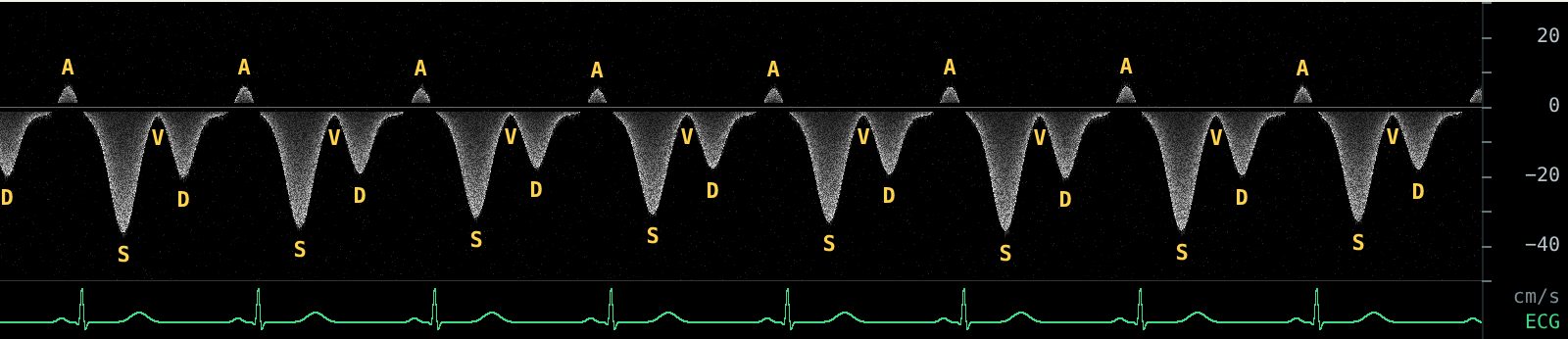
Normal: S larger than D. Both waves are below the baseline and S is the deeper one. The right atrium is filling at low pressure during systole.
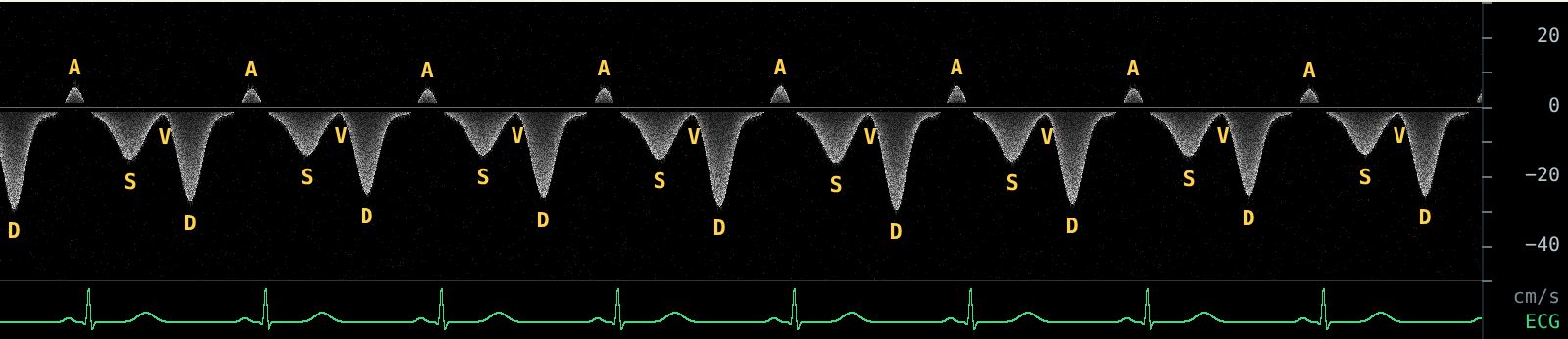
Mildly abnormal: S smaller than D. Both are still toward the heart, but rising right atrial pressure blunts the systolic suction, so D becomes the dominant wave.
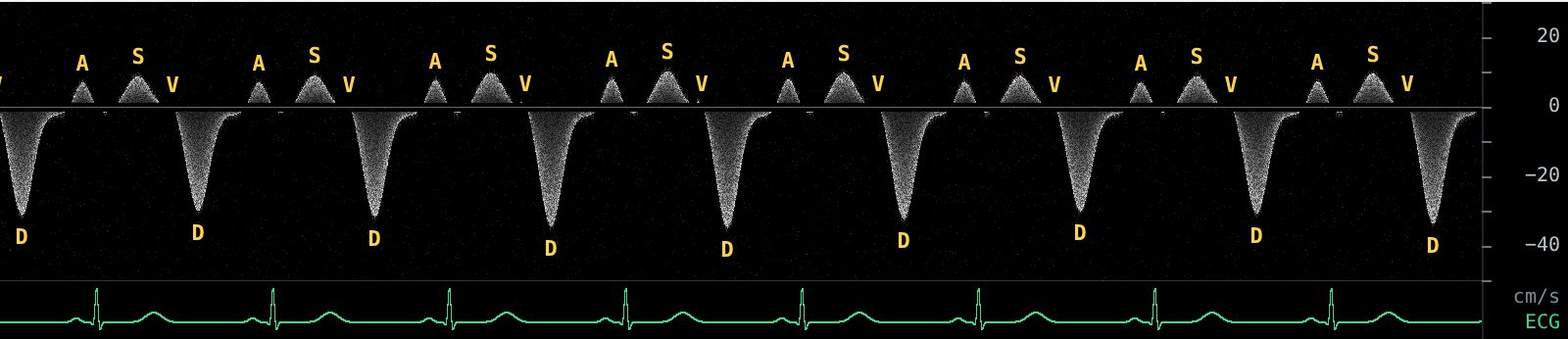
Severely abnormal: S reversed. The systolic wave flips above the baseline. Blood is moving back into the liver during systole. This is the pattern VExUS counts as severe (Beaubien-Souligny 2020).
Pitfalls
- Atrial fibrillation. There is no A wave, and S is often smaller than D even without congestion, because there is no atrial relaxation to help systolic filling. Interpret S less than D in AF with caution (Rola 2021).
- Tricuspid regurgitation. Severe TR reverses S by itself, regardless of volume. The pattern still means high systolic pressure in the right atrium, but the cause may be the valve.
- Cirrhosis and liver disease. A stiff liver dampens the hepatic vein waveform and can make it look flat or monophasic (Scheinfeld 2009).
- Respiration and mechanical ventilation. Waveforms change through the respiratory cycle. Look at several beats and grade the typical one.
Hepatic vein Doppler is one of three Doppler views in VExUS. It only counts once the IVC is 2 cm or wider. See the IVC guide and the full VExUS grading guide.
Practice reading these waveforms. The Venous Congestion Trainer plays live hepatic, portal and intrarenal Doppler with an ECG, lets you change each pattern and the IVC, and quizzes you on random cases with an answer key.
Related guides
References
- Beaubien-Souligny W, Rola P, Haycock K, et al. Quantifying systemic congestion with point-of-care ultrasound: development of the venous excess ultrasound grading system. Ultrasound J. 2020;12:16.
- Scheinfeld MH, Bilali A, Koenigsberg M. Understanding the spectral Doppler waveform of the hepatic veins in health and disease. Radiographics. 2009;29(7):2081-2098.
- Rola P, Miralles-Aguiar F, Argaiz E, et al. Clinical applications of the venous excess ultrasound (VExUS) score: conceptual review and case series. Ultrasound J. 2021;13:32.
- Argaiz ER, Koratala A, Reisinger N. Comprehensive assessment of fluid status by point-of-care ultrasonography. Kidney360. 2021;2(8):1326-1338.