Portal vein pulsatility
Normal portal venous flow is steady, because the liver sinusoids sit between the portal vein and the heart. When right-sided pressure gets high enough, the cardiac pulse is transmitted backward through the liver and the portal waveform starts to pulse.
Getting the trace
- Find the main portal vein at the liver hilum. A right intercostal view along the midaxillary line usually lines it up well with the beam.
- Put the pulsed wave gate in the main portal vein or the right portal branch. Portal flow toward the liver plots above the baseline.
- Set the scale low (around 30 to 40 cm/s) so the waveform fills the screen.
The formula
Vmax is the highest velocity in the cardiac cycle and Vmin is the lowest. Because it is a ratio, the Doppler angle cancels out, which makes it more forgiving than most Doppler measurements.
VExUS cutoffs
| Pulsatility fraction | VExUS pattern |
|---|---|
| Under 30% | Normal |
| 30 to 49% | Mildly abnormal |
| 50% or more | Severely abnormal |
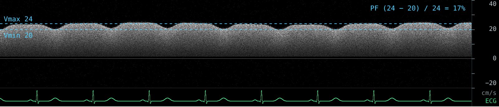
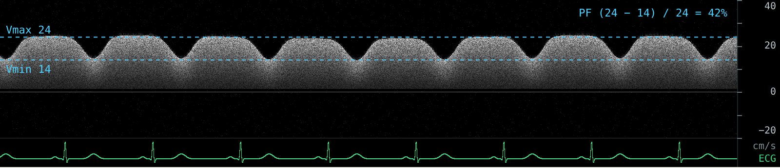
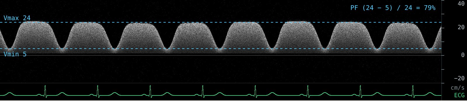
Flow reversal
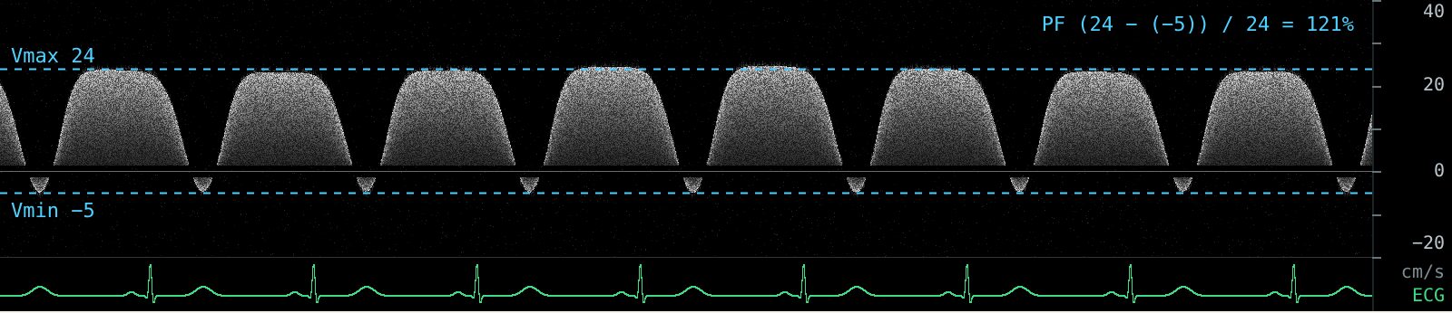
When Vmin drops below zero, portal flow briefly reverses with each beat. Plug the negative value into the formula and the fraction goes above 100%. It is still graded as severe.
Pitfalls
- Thin, healthy people can have some portal pulsatility without congestion. The IVC gate in VExUS helps here: a small IVC means grade 0 regardless (Rola 2021).
- Cirrhosis and portal hypertension dampen portal pulsatility and can reverse flow for reasons unrelated to the heart, so the portal vein is less useful in these patients.
- Breathing. Measure Vmax and Vmin within the same cardiac cycle and avoid beats distorted by a deep breath.
In a general ICU cohort, abnormal portal and hepatic Doppler patterns were associated with major adverse kidney events (Spiegel 2020). See the evidence summary.
Practice reading these waveforms. The Venous Congestion Trainer plays live hepatic, portal and intrarenal Doppler with an ECG, lets you change each pattern and the IVC, and quizzes you on random cases with an answer key.
Related guides
References
- Beaubien-Souligny W, Rola P, Haycock K, et al. Quantifying systemic congestion with point-of-care ultrasound: development of the venous excess ultrasound grading system. Ultrasound J. 2020;12:16.
- Rola P, Miralles-Aguiar F, Argaiz E, et al. Clinical applications of the venous excess ultrasound (VExUS) score: conceptual review and case series. Ultrasound J. 2021;13:32.
- Argaiz ER, Koratala A, Reisinger N. Comprehensive assessment of fluid status by point-of-care ultrasonography. Kidney360. 2021;2(8):1326-1338.
- Spiegel R, Teeter W, Sullivan S, et al. The use of venous Doppler to predict adverse kidney events in a general ICU cohort. Crit Care. 2020;24:615.