Intrarenal venous Doppler
The kidney is encapsulated, so when venous pressure rises, the renal veins lose the steady flow they normally have. Intrarenal venous Doppler shows that change directly, and it is the third Doppler view in VExUS.
Getting the trace
- Find the kidney in long axis from the flank. The right kidney is usually easier, through the liver.
- Turn on color Doppler with a low velocity scale and find the interlobar vessels between the medullary pyramids.
- Put a slightly wide pulsed wave gate over an artery and vein running together. The artery plots above the baseline and the vein below.
- Set the scale low (around 15 to 20 cm/s) and the wall filter low, or the slow venous signal disappears. Keep the ECG on to time the waves.
The three patterns
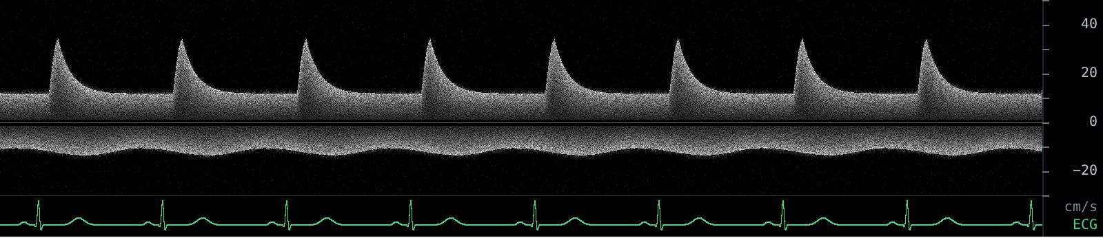
Normal: continuous. The venous signal below the baseline is steady through the whole cardiac cycle.
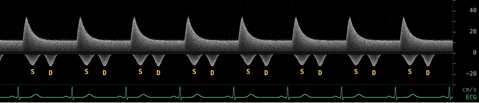
Mildly abnormal: biphasic. The venous flow breaks into two separate waves, one in systole (S) and one in diastole (D), with gaps between them.
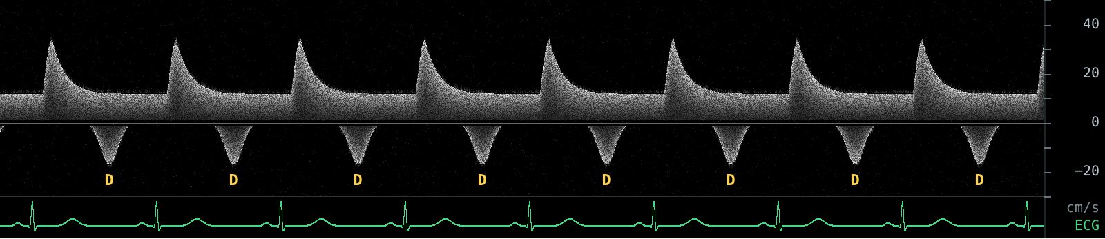
Severely abnormal: monophasic. Only the diastolic wave is left. Venous flow out of the kidney happens only when the tricuspid valve is open.
What the patterns mean
In heart failure patients, discontinuous intrarenal venous flow was associated with higher right atrial pressure and with worse outcomes, and the monophasic pattern carried the worst prognosis (Iida 2016). In VExUS, monophasic flow counts as a severe pattern (Beaubien-Souligny 2020).
Pitfalls
- Settings. A scale set for arterial flow or a high wall filter can make continuous venous flow look absent. Turn both down before calling a pattern.
- Chronic kidney disease and intrinsic renal disease change renal vascular resistance and can alter the venous waveform independent of congestion.
- Hard windows. Obesity, bowel gas and a patient who cannot hold their breath make this the hardest VExUS view. It is reasonable to report it as not obtained rather than guess.
More: hepatic vein Doppler, portal pulsatility and the full VExUS grading guide.
Practice reading these waveforms. The Venous Congestion Trainer plays live hepatic, portal and intrarenal Doppler with an ECG, lets you change each pattern and the IVC, and quizzes you on random cases with an answer key.
Related guides
References
- Beaubien-Souligny W, Rola P, Haycock K, et al. Quantifying systemic congestion with point-of-care ultrasound: development of the venous excess ultrasound grading system. Ultrasound J. 2020;12:16.
- Iida N, Seo Y, Sai S, et al. Clinical implications of intrarenal hemodynamic evaluation by Doppler ultrasonography in heart failure. JACC Heart Fail. 2016;4(8):674-682.
- Rola P, Miralles-Aguiar F, Argaiz E, et al. Clinical applications of the venous excess ultrasound (VExUS) score: conceptual review and case series. Ultrasound J. 2021;13:32.
- Argaiz ER, Koratala A, Reisinger N. Comprehensive assessment of fluid status by point-of-care ultrasonography. Kidney360. 2021;2(8):1326-1338.