IVC ultrasound for VExUS
VExUS starts with the inferior vena cava. If it is under 2 cm, the grade is 0 and the Doppler views are not scored. If it is 2 cm or more, you go on to the hepatic, portal and intrarenal veins.
Where to measure
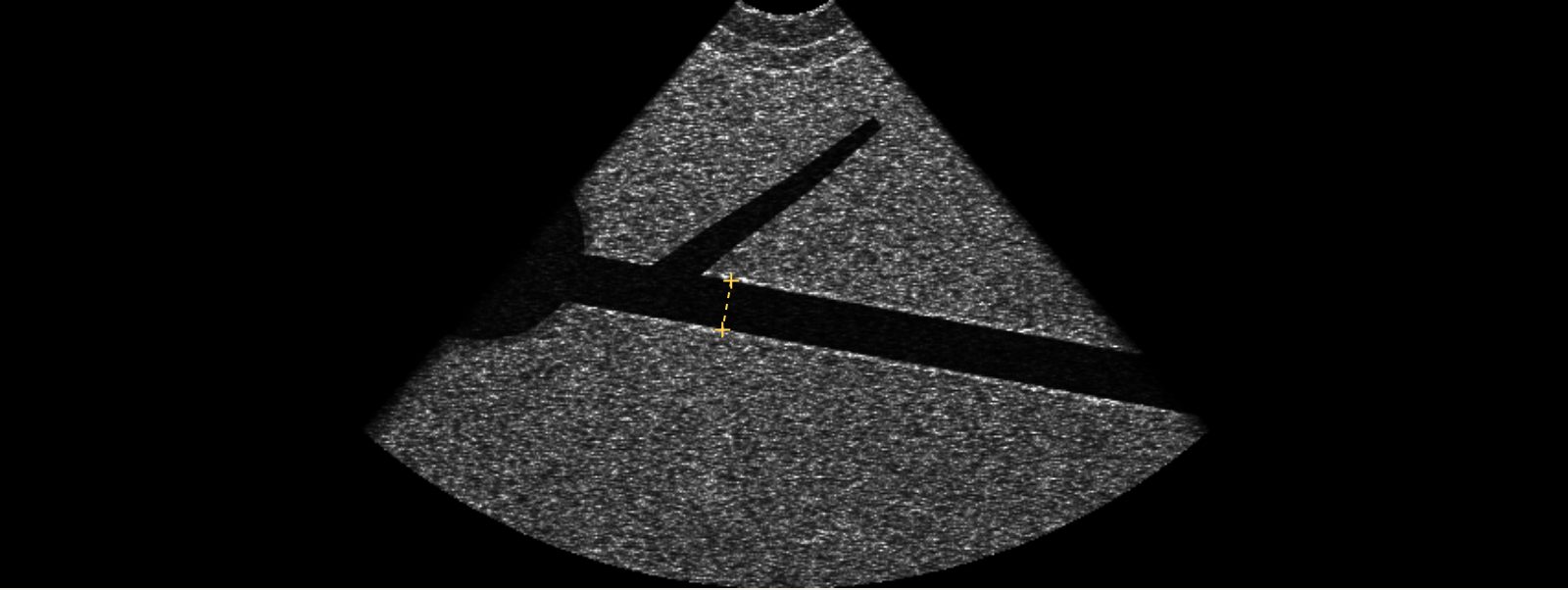
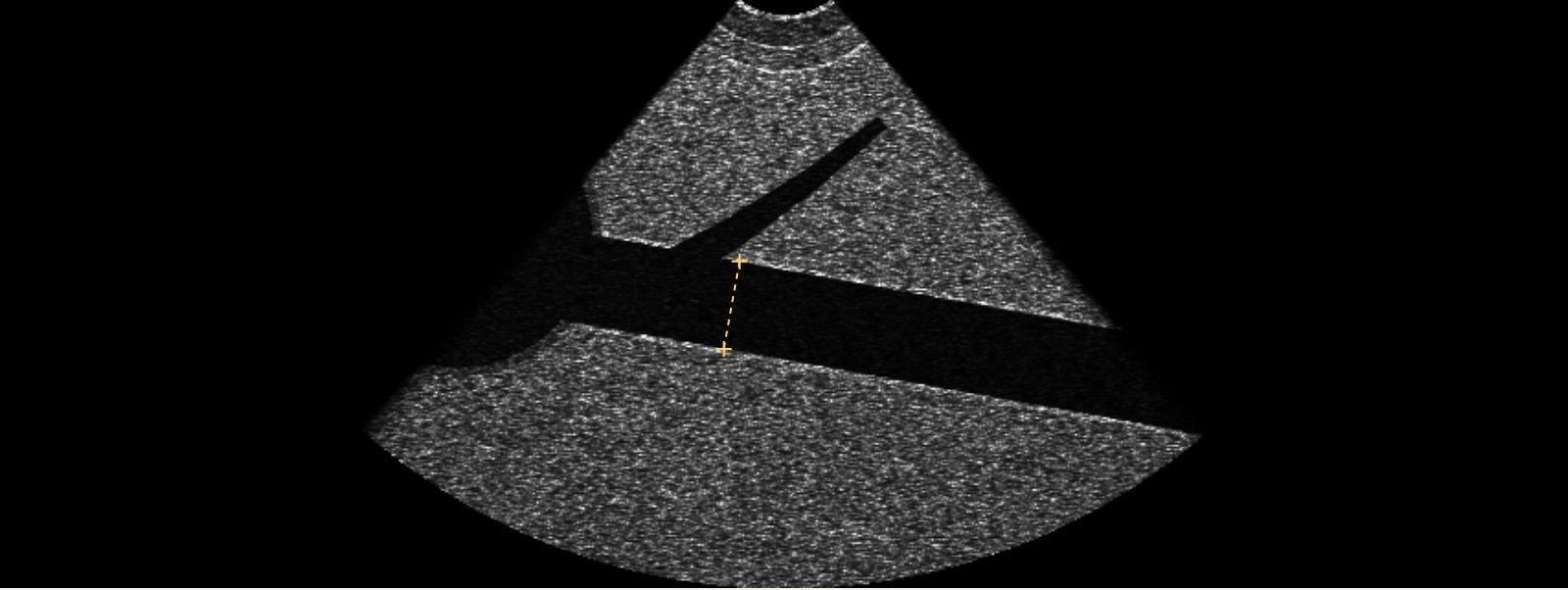
- Subcostal view with the probe marker toward the head. Find the IVC in long axis entering the right atrium, with the liver in front of it.
- Measure the diameter perpendicular to the vessel, about 1 to 2 cm from the right atrial junction, or just beyond where the hepatic veins join.
- Measure at its widest point in the respiratory cycle (end expiration in a spontaneously breathing patient).
The 2 cm cutoff
A plethoric IVC is necessary but not sufficient for systemic venous congestion. In VExUS it works as a gate: below 2 cm the patient is grade 0, and only at 2 cm or more do the Doppler patterns decide grades 1 to 3 (Beaubien-Souligny 2020).
For estimating right atrial pressure, the ASE uses 2.1 cm and 50% collapse with a sniff: under 2.1 cm and collapsing more than 50% suggests about 3 mmHg; over 2.1 cm and collapsing less than 50% suggests about 15 mmHg (Rudski 2010).
| Grade | IVC | Hepatic, portal and intrarenal Doppler |
|---|---|---|
| 0, no congestion | Under 2 cm | Not graded |
| 1, mild | 2 cm or more | Any mix of normal and mildly abnormal patterns |
| 2, moderate | 2 cm or more | One severely abnormal pattern |
| 3, severe | 2 cm or more | Two or three severely abnormal patterns |
Pitfalls
- Off-axis cuts. If the beam slices the side of the IVC instead of its center, the vessel looks smaller than it is. Fan through it, or check it in short axis.
- Mechanical ventilation raises intrathoracic pressure and often dilates the IVC without congestion. Collapsibility rules for spontaneous breathing do not apply.
- Athletes and tall patients can have a large IVC at normal pressures.
- Raised intra-abdominal pressure can compress the IVC and make it look small even when right-sided pressure is high (Rola 2021).
- Mistaking the aorta. The aorta is thick-walled, pulsatile and does not join the right atrium. Follow the vessel into the heart.
Practice reading these waveforms. The Venous Congestion Trainer plays live hepatic, portal and intrarenal Doppler with an ECG, lets you change each pattern and the IVC, and quizzes you on random cases with an answer key.
Related guides
References
- Beaubien-Souligny W, Rola P, Haycock K, et al. Quantifying systemic congestion with point-of-care ultrasound: development of the venous excess ultrasound grading system. Ultrasound J. 2020;12:16.
- Rudski LG, Lai WW, Afilalo J, et al. Guidelines for the echocardiographic assessment of the right heart in adults. J Am Soc Echocardiogr. 2010;23(7):685-713.
- Rola P, Miralles-Aguiar F, Argaiz E, et al. Clinical applications of the venous excess ultrasound (VExUS) score: conceptual review and case series. Ultrasound J. 2021;13:32.
- Argaiz ER, Koratala A, Reisinger N. Comprehensive assessment of fluid status by point-of-care ultrasonography. Kidney360. 2021;2(8):1326-1338.